2019 Volume No 37 – pages 420-430
Title: A metaphyseal fracture rat model for mechanistic studies of osteoporotic bone healing |
Authors: RMY Wong, U Thormann, MHV Choy, YN Chim, MCM Li, JY Wang, KS Leung, JCY Cheng, V Alt, SKH Chow, WH Cheung |
Address: Department of Orthopaedics and Traumatology, Prince of Wales Hospital, The Chinese University of Hong Kong, Shatin, New Territories, Hong Kong, China. |
E-mail: skhchow at ort.cuhk.edu.hk |
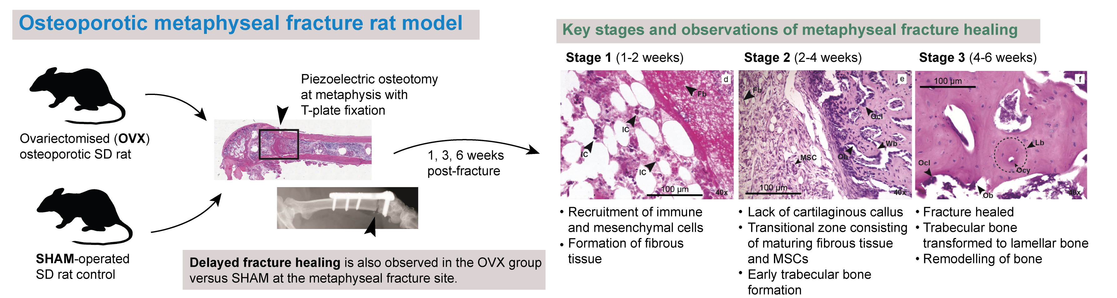
Abstract: Most osteoporotic fractures occur at metaphyseal regions of long bones. The present study proposed a clinically relevant animal model that satisfied: i) induction of osteoporosis, ii) unilateral complete osteotomy at metaphysis, iii) internal fixation. 6 months old female Sprague-Dawley rats (n = 64) were randomly divided into the ovariectomised-metaphyseal osteotomy (OVX, n = 32) and metaphyseal osteotomy (SHAM, n = 32) groups. The metaphyseal-osteotomy model was created with a plate-fixation of the osteotomy and assessed by X-ray, micro-computed tomography, histomorphometry and mechanical testing at weeks 1, 3 and 6. X-ray results showed complete healing of metaphyseal osteotomy at week 6. Histology showed 3 stages of metaphyseal healing. Stage 1 was characterised by fibrous tissue, consisting of disorganised orientation of collagen fibres, and infiltration of immune cells. At stage 2, a transitional zone consisting of maturing fibrous tissue and differentiating mesenchymal cells with early trabecular bone formation and disorganised woven bone were observed. During stage 3, cortical bone ends unified and woven bone underwent transformation to lamellar bone. OVX group healing was significantly delayed when compared to SHAM samples. |
Key Words: Osteoporotic fracture healing, metaphyseal fracture, intramembranous ossification, endochondral ossification. |
Publication date: May 22nd 2019 |
Article download: Pages
420-430 (PDF file)
|